What is CBCT scan : 5 Advantages and Disadvantages
Advertisement
CBCT (Cone Beam Computed Tomography) is a specialized imaging technique commonly used in dentistry and maxillofacial applications to generate detailed 3D images. Advantages include lower radiation exposure than conventional CT and high imaging accuracy, while disadvantages include limited soft tissue visualization and higher equipment costs.
Conventional Radiography: This technique generates a two-dimensional image by superimposing images from successive layers of the body in the path of X-rays. A key challenge is that the image of one layer is obscured by the superimposition of images from layers above and below it.
Computed Tomography (CT): CT addresses the limitations of conventional radiography by recording images of selected layers sharply while recording images of other layers unsharply. Tomography involves synchronized movement of any two of the following three subjects: the X-ray tube, the film, and the patient, while the third remains stationary. The most common and widely used method involves keeping the patient stationary while the X-ray tube and film (or detector) move in coordination.
What is Cone Beam Computed Tomography (CBCT)?
CBCT is a variation of traditional computed tomography. Unlike a CT scanner, it uses an X-ray tube and a detector panel that rotates around the patient, capturing data in a cone-shaped X-ray beam instead of the slices captured by CT. Algorithms reconstruct these images to generate high-resolution, three-dimensional images.
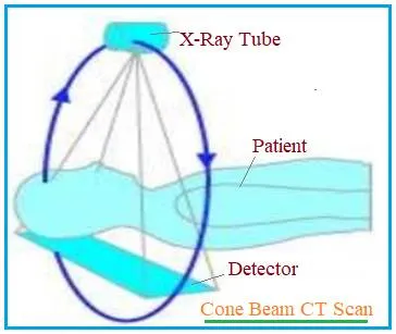
Let’s explore how CBCT works. All CBCT scanners use an X-ray source and detector mounted on a rotating gantry. During the gantry’s rotation, the X-ray source emits divergent cone-shaped radiation, and the receptor records the residual X-rays after they’ve been attenuated by the patient’s tissues.
The X-ray source and detector move through an arc of 180 to 360 degrees to produce multiple planar projection images. These images constitute the raw primary data. An algorithm running on a computer reconstructs this data to generate cross-sectional images. Sensors such as image sensors (PSP - Photo Stimulable Phosphorus Plates), CCD sensors, and FPD (Flat Panel Detector) are used for X-ray detection.
Advantages of CBCT Scan
- Rapid Scan Time: CBCT offers significantly faster scan times.
- Reduced Radiation Exposure: Patients experience less radiation exposure compared to CT scans.
- Three-Dimensional Volume Rendering: CBCT delivers detailed three-dimensional volume renderings.
- Accurate Images with Good Spatial Resolution: CBCT provides accurate images with excellent spatial resolution.
- Economical, Comfortable, and Safe: CBCT is an economical, comfortable, and safe imaging technique.
- Interactive Display Modes and Multiplanar Reformatting: CBCT supports interactive display modes and multiplanar reformatting, offering flexibility in image analysis.
- Improved Treatment Planning: CBCT imaging provides insights into treatment planning that are not achievable with other methods, enabling clinicians to provide more predictable patient care.
Disadvantages of Cone Beam Computed Tomography (CBCT) scan
- Poor Contrast Resolution: CBCT offers poor contrast resolution, making it difficult to visualize soft tissues effectively.
- Artifacts: Certain artifacts cannot be resolved by CBCT and may appear in the image.
- Image Noise: The images generated by CBCT can exhibit noise.
- Not Always Necessary: CBCT is not required when 2D imaging is sufficient.
- Higher Radiation: Although CBCT delivers less radiation than conventional CT, it still exposes patients to a higher dose compared to standard 2D dental radiographs, which may be a concern for frequent or unnecessary use.
Summary
Cone Beam Computed Tomography (CBCT) scan is a valuable imaging technique that provides detailed 3D views, especially useful in dental, maxillofacial and ENT applications. It offers improved spatial resolution and precise anatomical visualization compared to traditional 2D imaging. However, its limitations such as poor soft tissue contrast, image artifacts, noise, and higher radiation exposure than 2D methods; must be carefully considered. When used appropriately, CBCT significantly enhances diagnostic accuracy and treatment planning in targeted clinical scenarios.